BBB

Pial arteries rest on the glia limitans, which envelops the brain surface. Penetrating arterioles are surrounded by astrocytic endfeet that express several specialized proteins. Astrocytes have fine processes in close proximity to synapses and occupy nonoverlapping spatial domains. Aqp4, aquaporin 4.
Glymphatics
To discover underlying mechanisms of waste clearance in the brain, researchers have implanted a window into the skull of mice and used two-photon imaging techniques that allow them to directly observe the movemement of cerebrospinal fluid (CSF) through the living brain!

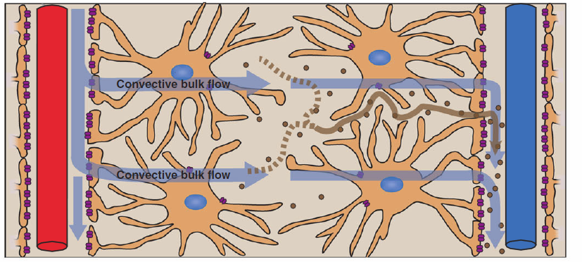
The researchers found that the brain itself is surrounded by a membrane called the arachnoid and bathed in cerebral spinal fluid (CSF). CSF flows into the interior of the brain through the same pathways as the arteries that carry blood and is drawn into brain tissue via a system of conduits that are controlled by astrocytes ( a type of glia). The CSF is flushed through the brain tissue at high speeds, which sweeps away excess “garbage” that might be located in the brain. The fluid and waste are exchanged with a similar system that parallels veins which carries the waste out of the brain and down the spine where it is eventually transferred to the lymphatic system and from there to the liver, where it is ultimately broken down.The scientists decided to coin this newly discovered system the “glymphatic system.”
Convective glymphatic fluxes of CSF and ISF propel the waste products of neuron metabolism into the paravenous space, from which they are directed into lymphatic vessels and ultimately return to the general circulation for clearance by the kidney and liver.
CREDIT: K. SUTLIFF/SCIENCE
Virchow-Robin spaces (VRS)
Virchow–Robin spaces (VRS) are perivascular, fluid-filled canals that surround perforating arteries and veins in the parenchyma of the brain. VRS are extremely small and can usually only be seen on MR images when dilated. While many normal brains will show a few dilated VRS, an increase in dilated VRS has been shown to correlate with the incidence of several neurodegenerative diseases, making the spaces a popular topic of research.[1]

Virchow–Robin spaces are gaps containing interstitial fluid that span between blood vessels and the brain matter which they penetrate.[4] Like the blood vessels around which they form, Virchow–Robin spaces are found in both the subarachnoid space and the subpial space.[5] VRS surrounding arteries in the cerebral cortex and the basal ganglia are separated from the subpial space by one or two layers of leptomeninges, respectively, as well as the pia mater.[2] By virtue of the leptomeningeal cell layer, VRS belonging to the subarachnoid space are continuous with VRS of the subpial space. The direct communication between VRS of the subarachnoid space and the subpial space is unique to the brain’s arteries, as no leptomeningeal layers surround the brain’s veins.[2][5] Use of the scanning electron microscope has determined that VRS surrounding blood vessels in the subarachnoid space are not continuous with the subarachnoid space because of the presence of pia mater cells joined by desmosomes.[6]
Virchow–Robin spaces may be enlarged to a diameter of five millimeters in healthy humans and are usually harmless. When enlarged, they can disrupt the function of the brain regions into which they project.[4]Dilation can occur on one or both sides of the brain.[2]
 |
2014년 9월 29일 월요일
Glymphatics and virchow-robin space
Glia and excitability in epilepsy and infantile spasm

Figure 1. Schematic model depicting selected interactions between astrocytes and excitatory neurons. Voltage-gated Na+ and K+ channels (1) generate action potentials in the presynaptic neuron, leading to the exocytotic synaptic release of neurotransmitter glutamate(2). Glutamate activates AMPA and NMDA receptors (3) in the postsynaptic membrane, causing excitatory synaptic potentials generated by influx of Na+ and Ca2+. If sufficiently strong, synaptic excitation leads to epileptiform discharges (4). Glutamate is taken up into reactive astrocytes by the EAAT1 (GLAST) and EAAT2 (GLT-1) transporters (5) and is converted to glutamine by glutamine synthetase (6). Glutamine is a substrate for the production of GABA in inhibitory GABAergic neurons (not shown). Loss of glutamine synthetase in reactive astrocytes leads to a decrease in GABA production. K+ released from neurons by voltage-gated (outwardly rectifying) K+ channels enters astrocytes via inwardly rectifying K+ channels (Kir4.1) (7) and is distributed into capillaries. Aquaporin-4 (AQP4) concentrated at astrocytic endfoot processes regulates water balance (8). Ca2+ waves (9) stimulate the release of gliotransmitters (10) that can influence neuronal excitability. The inhibitory substance adenosine is taken up into astrocytes by the equilibrative nucleoside transporters ENT1 and ENT2 and concentrative nucleoside transporter CNT2. Excessive adenosine kinase in reactive astrocytes increases the removal of adenosine (11), enhancing hyperexcitability.
Categories
1. AP --> glutamate release --> AMPA/NMDA --> Na, Ca influx
; epileptiform discharge, if strong
2. Glutamate uptake (EAAT1,2) in astrocyte --> glutamine --> GABA production by GS
; epileptiform discharge, if EAAT1,2 mutation, GS deficient
3. K release from neuron by voltage-gated K channel à inward K channel (Kir4.1) in astrocyte
; reduced Kir4.1 expression --> epileptogenesis
4. AQP4 distribution in astrocyte
5. Ca wave in astrocyte --> gliotransmitter release
6. Adenosine in astrocyte (uptaken by ENT1,2) --> enhanced adenosine kinase (ADK) --> AMP
Glia and epilepsy
1. Water and K+ buffering
1.AQP4
2.Kir4.1
2. Regulating Neurotransmission
1.GLAST, GLT-1 (EAAT1, EAAT2)
2.ADK
3.GS
3. Gliotransmission
1. Ca wave by G-protein-coupled recetor-induced increased PLC or cyclooxygenase-2 prostaglandin signaling
2. D-serine
3. GABA
1.GLAST, GLT-1 (EAAT1, EAAT2)
2.ADK
3.GS
3. Gliotransmission
1. Ca wave by G-protein-coupled recetor-induced increased PLC or cyclooxygenase-2 prostaglandin signaling
2. D-serine
3. GABA
4. Vasculature and BBB
1. VEGF
2. Albumin-mediated activation of TGFb
3. p-glycoprotein
5. Glia-mediated immunity and inflammation

Table 1. Mechanisms of glia-mediated neuronal hyperexcitablity
1. VEGF
2. Albumin-mediated activation of TGFb
3. p-glycoprotein
5. Glia-mediated immunity and inflammation
Table 1. Mechanisms of glia-mediated neuronal hyperexcitablity
2014년 9월 25일 목요일
충남대학교 의학전문대학원 연구력 강화 워크샵 천안상록리조트 (2014. 9. 23)
Essential component for High Impact Journal!!
- Originality, Breakthrough
- Conceptual Advance
- Controversy
- Paradigm Shift


Graduate Student Yi won the Outstanding Young Scientist award.

Graduate student passed Prof Kim, the opponent defense in a swift move. OTL
2014년 9월 14일 일요일
2014 9 Japan Neuroscience Meeting in Yokohama
2014. 9. 11- 9.14 Pacifico Yokohama in Yokohama, Japan


Short talk presentation (Min-Hee Yi)

피드 구독하기:
글 (Atom)